Flexible endoscopy- Gastroscopy and Colonoscopy
Gastric Dilatation-Volvulus (GDV)
What is Bloat GDV?
Gastric Dilatation-Volvulus (GDV) is a rapidly progressive life-threatening condition that affects millions of dogs every year. In the beginning, the stomach fills with gas, food, foam, or air, causing the stomach to bloat, causing a gastric dilatation or enlargement. If the condition persists, the stomach continues to fill with gas and twists and progresses from simple gastric dilatation or Bloat to GDV. With no release, the stomach continues to expand and twist, cutting off the blood supply. At the same time, pressure is placed on internal organs, making it difficult for your dog to breathe and creating a life-threatening emergency where surgery and immediate veterinary care are needed.
The exact cause of Bloat GDV is unknown, but there is a multitude of factors that can make a dog more susceptible to developing this condition, such as:
- Genetics – Large breed dogs with deep, narrow chests can be at higher risks at developing GDV
- Eating habits – Eating one large meal a day, exercising after eating, and eating or drinking too quickly can be contributing factors
- Age – Dogs over 7 are more likely to develop GDV than others
It is important to note that any dog, regardless of age, size, and breed, can develop GDV.
SYMPTOMS
Most dogs will go into shock as soon as signs of GDV are realized, and death can occur within a matter of a few hours. It is essential to take your dog to the vet as soon as you notice any symptoms of GDV. The following are the most common signs of bloat GDV in dogs:
- Enlarged abdomen
- Rapid or shallow breathing
- Increase in heart rate
- Cold body temperature
- Unsuccessful attempts to vomit
- Excessive saliva
- Pale nose or gums
- Restlessness, pacing, and anxiety
- Shock
- Collapsing
TREATMENT
Bloat GDV is a life-threatening emergency that requires immediate veterinary attention. Once at the clinic, your vet will be able to relieve pressure on the stomach wall and internal organs. Shock treatment will also begin right away so that once your dog is stable, the surgical correction of GDV can begin if necessary.
WHAT CAN YOU DO?
Bloat GDV can be tough to prevent because the exact cause is not always known, and even with preventions, sometimes your dog may still get Bloat GDV. Some things you can do to help your dog avoid developing this condition include:
- Breaking up Meals – Instead of giving large meals, multiple meals throughout the day of smaller portions can help.
- Slow down their eating – overzealous eats may be at risk for bloat. Special bowls like puzzle feeders are designed to slow your dog’s intake. If your dog has siblings, separating them during meals can help them not feel rushed.
- Avoid exercising after a meal – encourage your dog to relax after eating.
- Keep water available at all times.
Recommend gastropexy surgery, where the stomach is attached to the body wall to reduce the likelihood of the dog’s stomach rotating. Please note that though this does prevent twisting of the stomach in most cases, there is still a chance of your dog’s stomach twisting and developing into GDV even with surgical preventions.
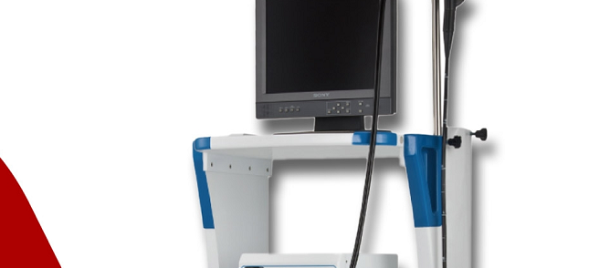
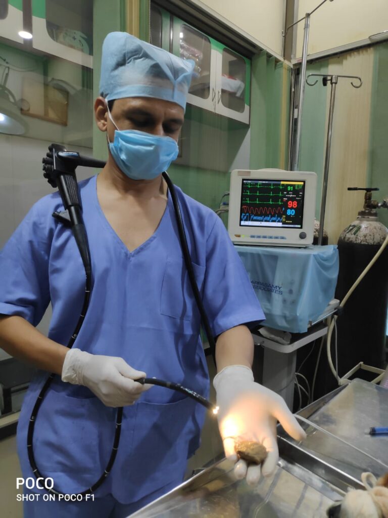
Endoscopy
At Small Animal Clinic, we have flexible video endoscopy system .This is to access your pets esophagus , stomach and small portion of duodenum ( small intestine) Colonoscopy involves accessing of colon and rectal area. Procedure is very simple and minimum invasive.
Your dog has been scheduled for an endoscopic examination. The purpose of this procedure is to make a diagnosis of the disease that has been causing your pet’s clinical signs of vomiting, diarrhea, weight loss, abdominal pain or swelling or loss of appetite.
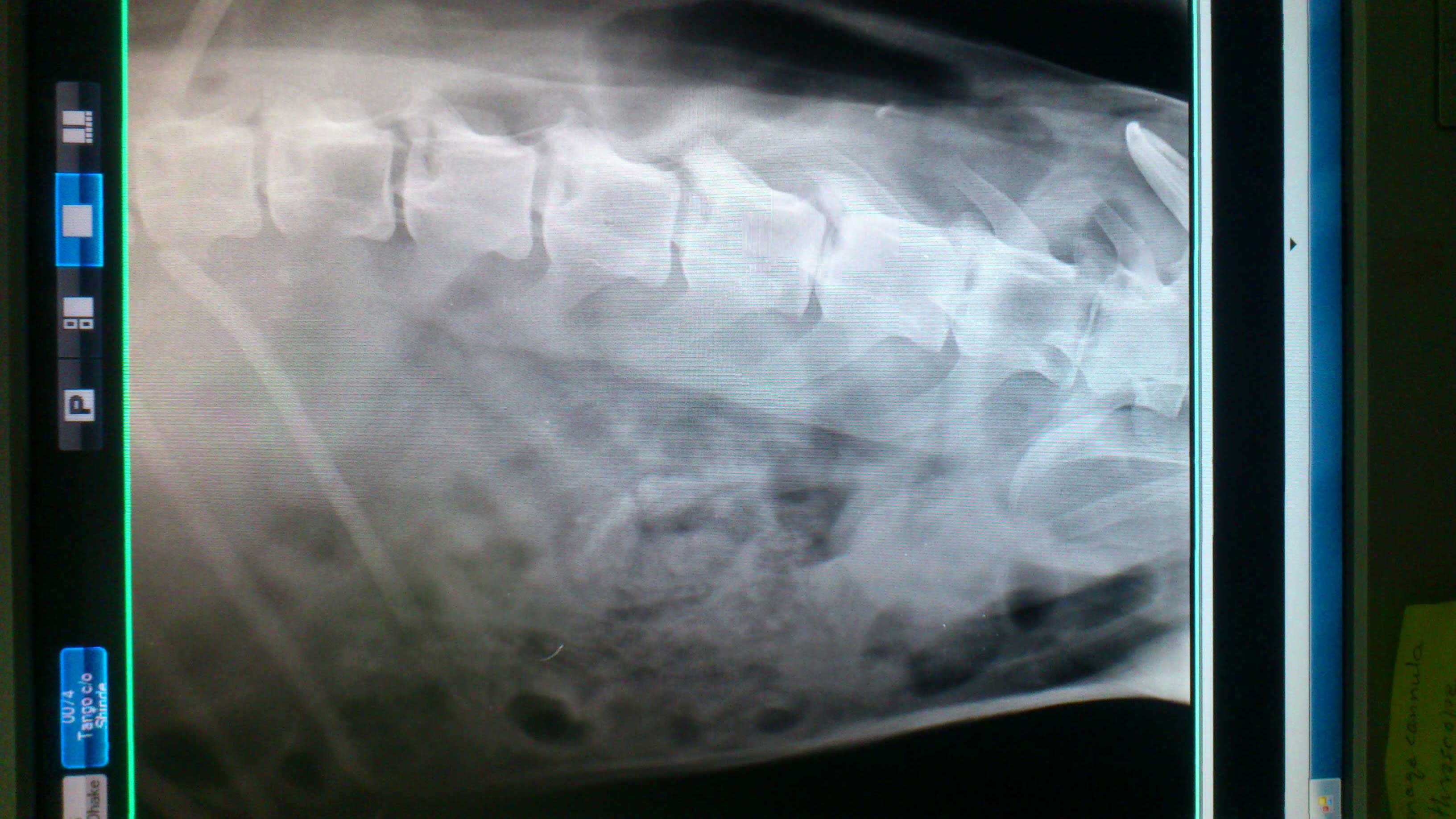
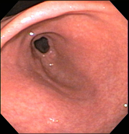
An endoscope is a flexible tube with a viewing port and a video camera attachment that is inserted either into the stomach through the mouth or the colon via the rectum. The endoscope permits inspection of the inside of these hollow organs. If the stomach is being examined, the esophagus can be inspected as the endoscope is being passed into the stomach.
The endoscope allows full color viewing of the esophagus, stomach and the upper part of the small intestine or the colon.
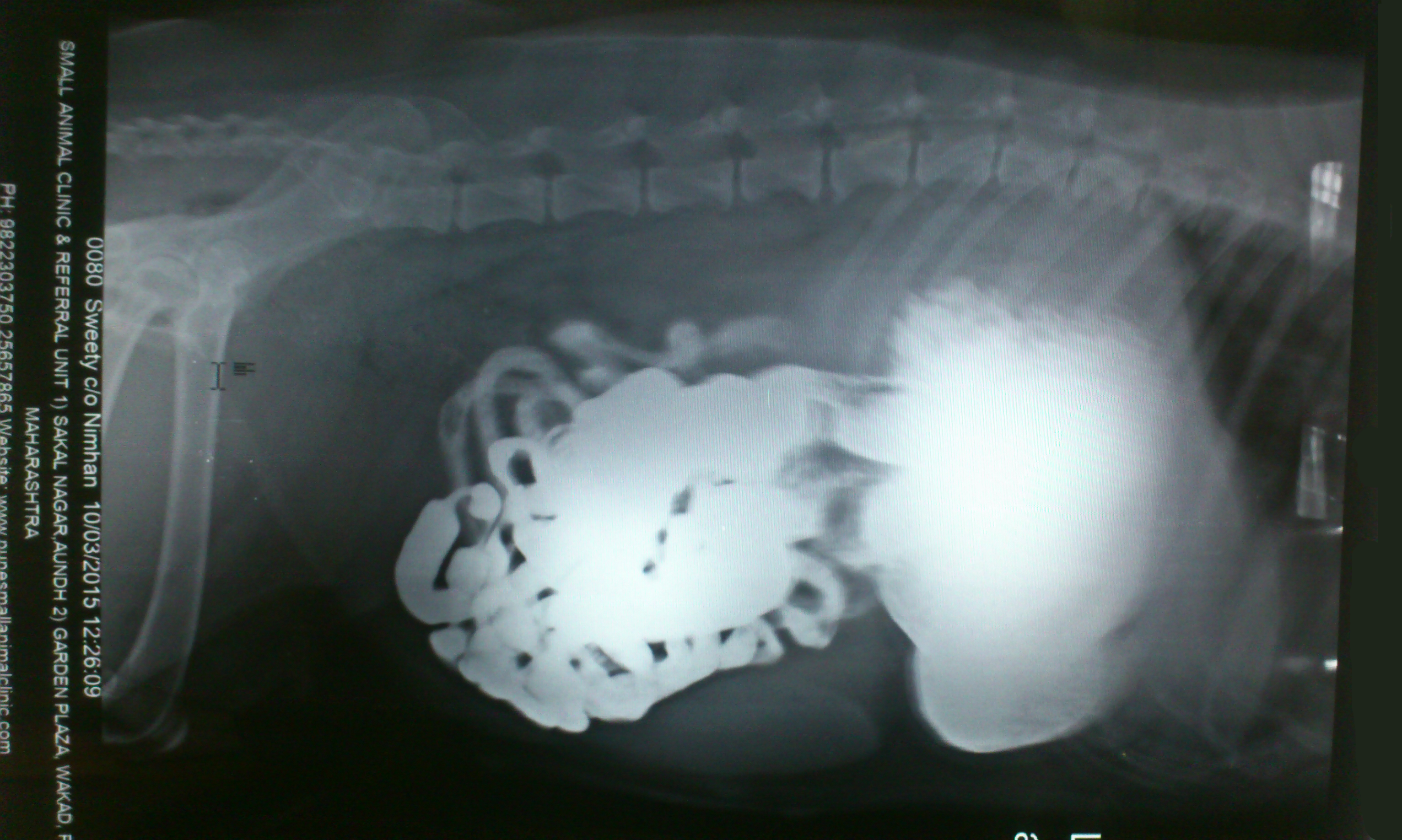
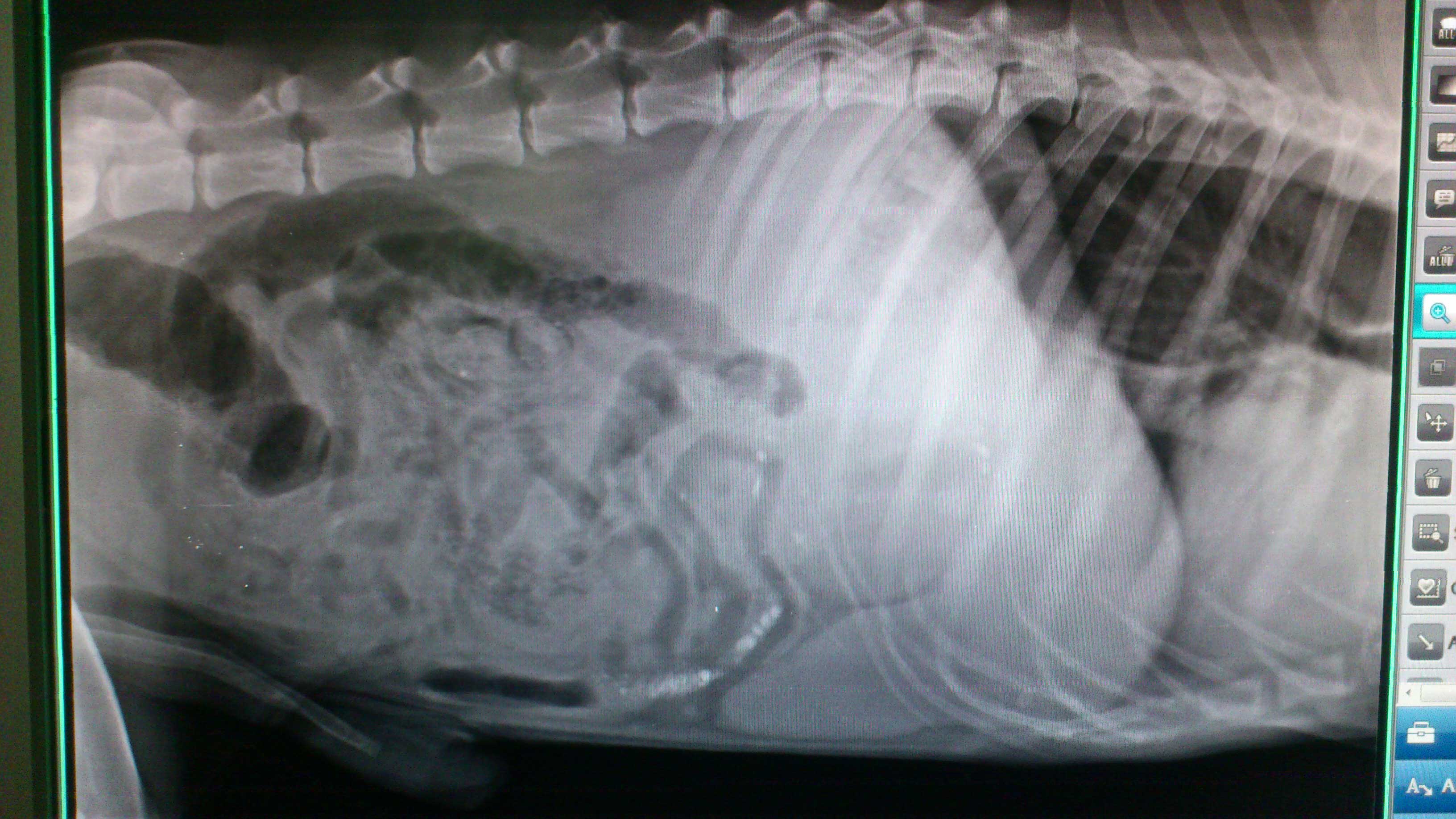
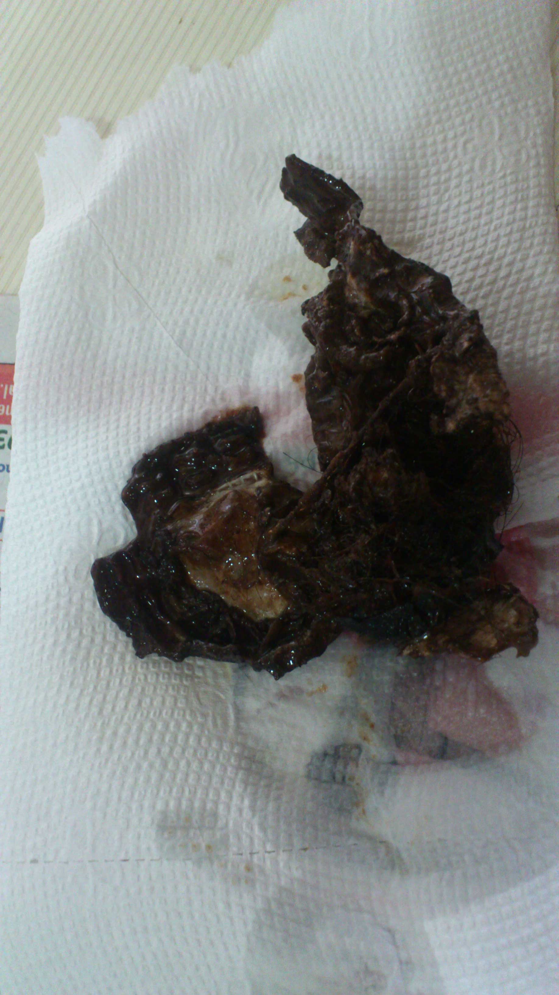
The examiner can identify abnormalities such as inflammation, abnormal swelling, or areas of scarring or stricture (abnormal narrowing). If a foreign body such as a bone, stick, rock, toy, coin, or hairball is seen, it can usually be seen and retrieved.
While seeing an abnormal lesion or suspicious area gives valuable information, it is usually necessary to biopsy the area in order to reach a diagnosis. The endoscope has a tiny channel through which a biopsy instrument can be passed. Precise biopsy samples can be taken of any abnormal areas and then submitted to a pathologist for microscopic evaluation.
In many cases we can diagnose cancer of the gastrointestinal tract using the endoscope. However, some tumors do not affect the mucosa or inner lining of the stomach or colon. Since the biopsy procedure only samples the mucosa it is possible to miss detecting a tumor that involves only the deeper layers of the intestinal tract. In these unusual cases, the biopsy results are normal yet the dog continues to experience clinical signs. In order to reach a diagnosis in these cases, full-thickness biopsies obtained through an exploratory surgery, (exploratory laparotomy) or non-invasive tests such as an MRI (Magnetic Resonance Imaging) may be required.
It is vital that the stomach and intestinal tract be empty of all food and fecal matter prior to an endoscopic evaluation. A complete twelve-hour fast is usually sufficient if the stomach is being examined. If the colon is to be examined, oral medication is begun twelve to eighteen hours before the procedure to remove fecal material from the entire intestinal tract. Fasting for twelve to eighteen hours is also necessary so that new fecal material does not form. On the morning of the procedure, one or more enemas are given to remove any remaining stool from the lower intestinal tract.
Is general anesthesia required?
“It is impossible to safely pass an endoscope into a conscious dog’s stomach or colon.”
Yes. It is impossible to safely pass an endoscope into a conscious dog’s stomach or colon. Most dogs will require only a short-acting anesthesia and the patient is allowed to go home shortly after completion of the procedure.Laboratory testing is the foundation of diagnostics in veterinary medicine. As a dedicated provider of quality veterinary care we provide our patients with both referral laboratory and on-site services. General laboratory samples are entrusted to the experts at our outside reference laboratory. However, rapid results are crucial for emergency care. Our in-house laboratory equipment provides us with the necessary information for treating our emergency patients promptly and running important pre-anesthetic panels. In addition, our on-site laboratory equipment enables us to continuously monitor critical patients’ blood values as needed
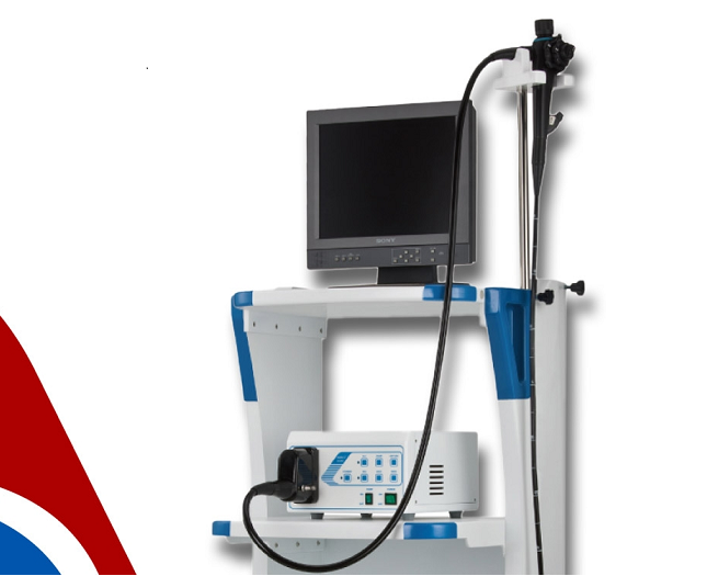
Endoscopy Machine
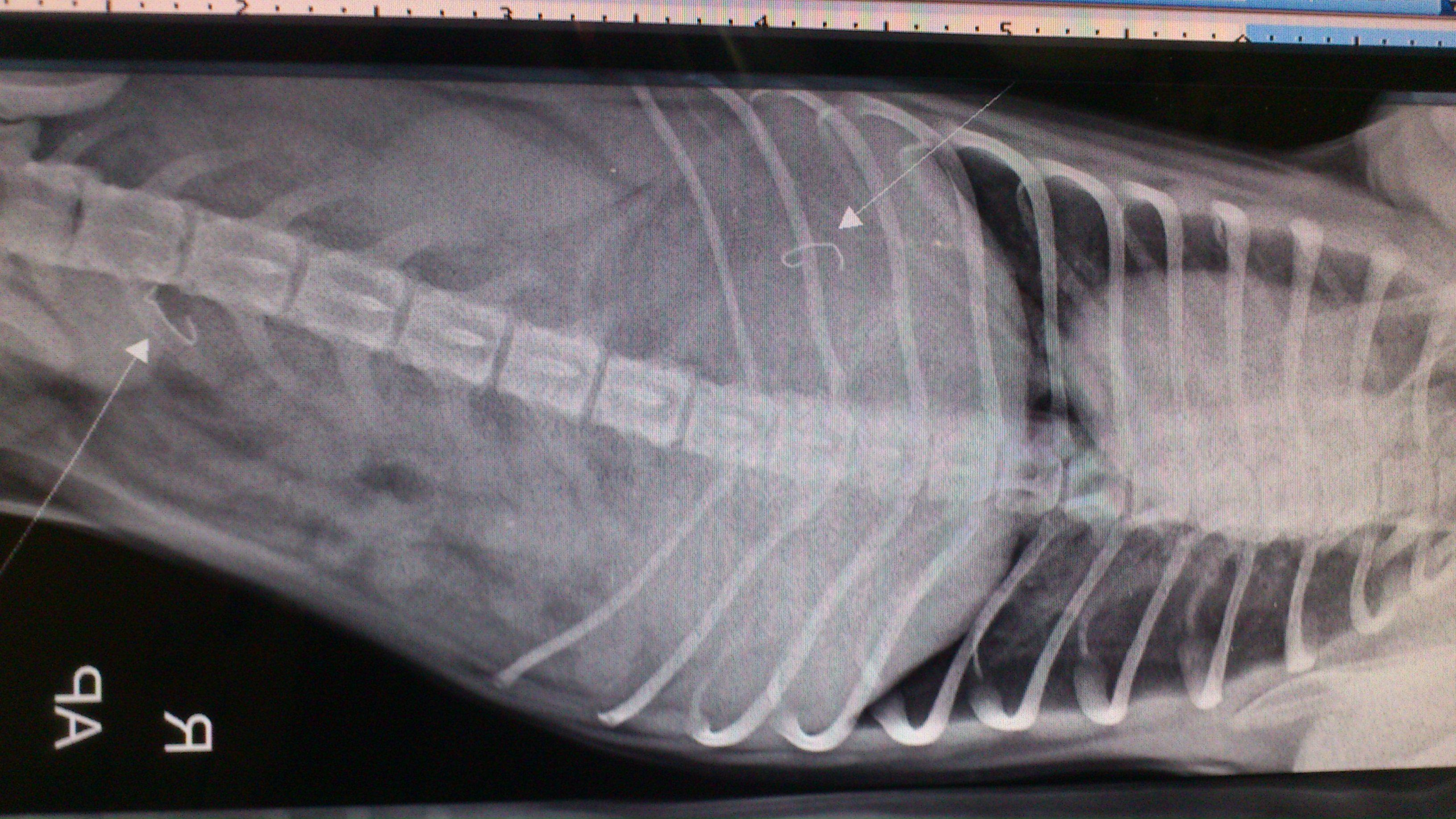
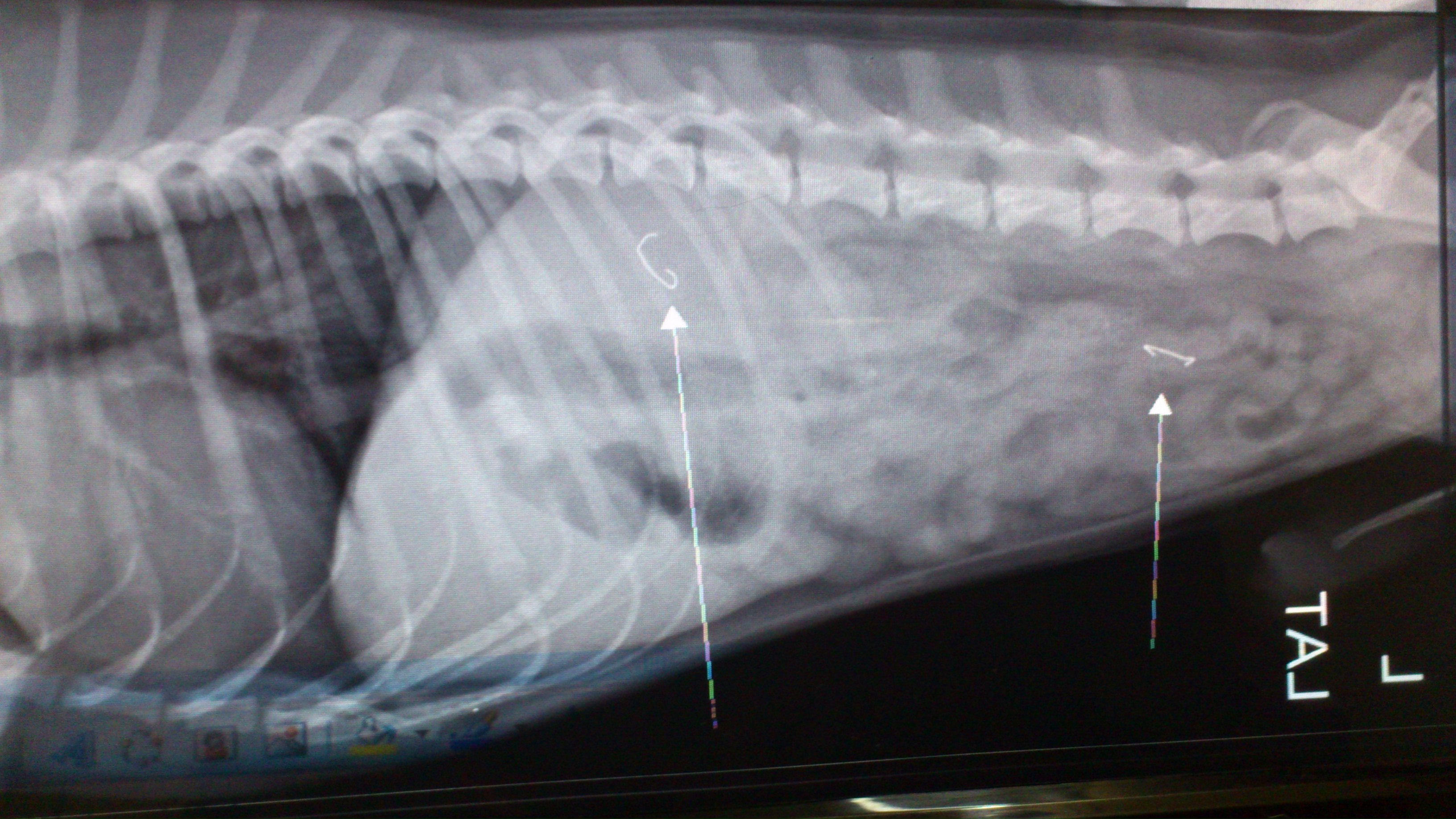
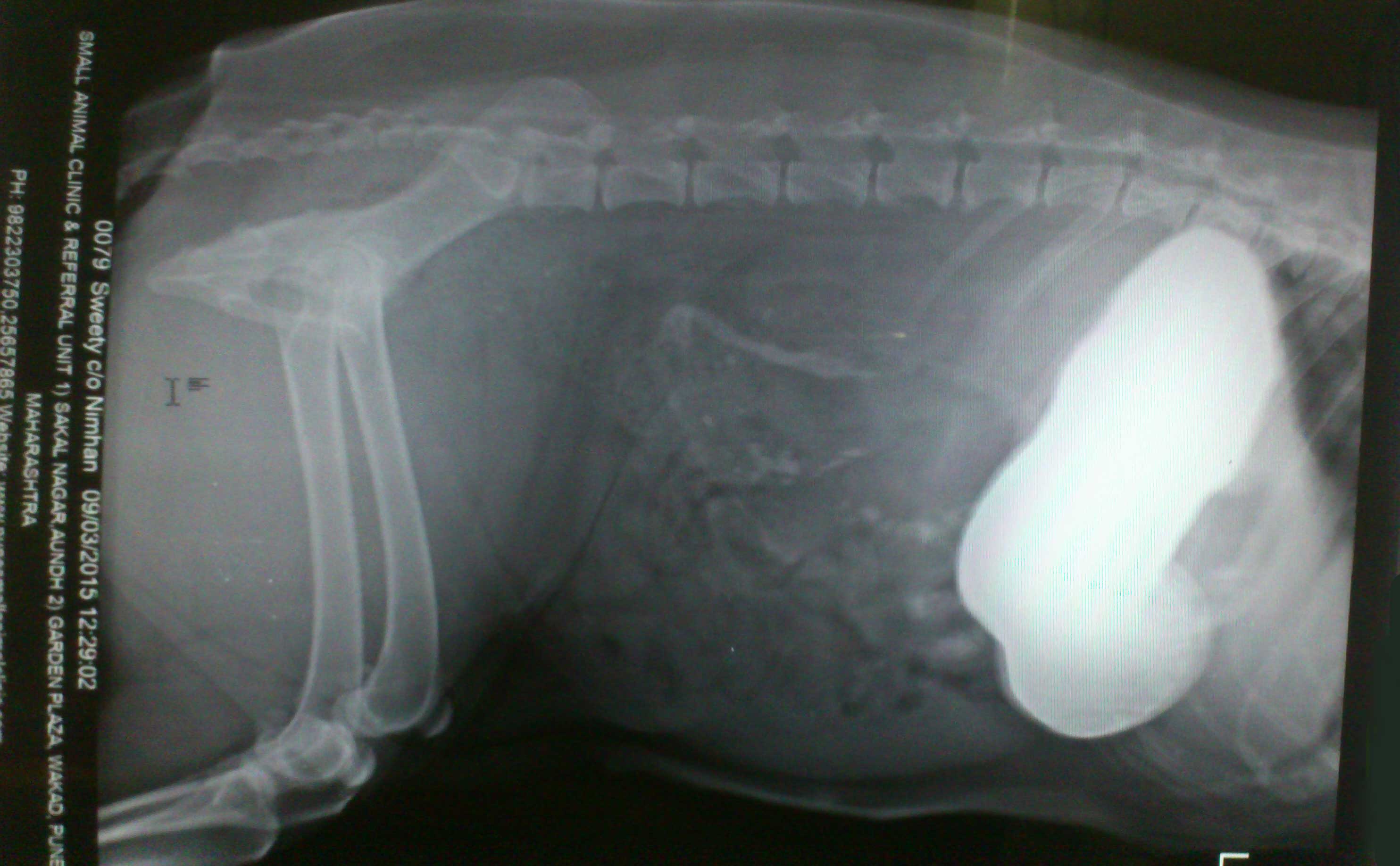
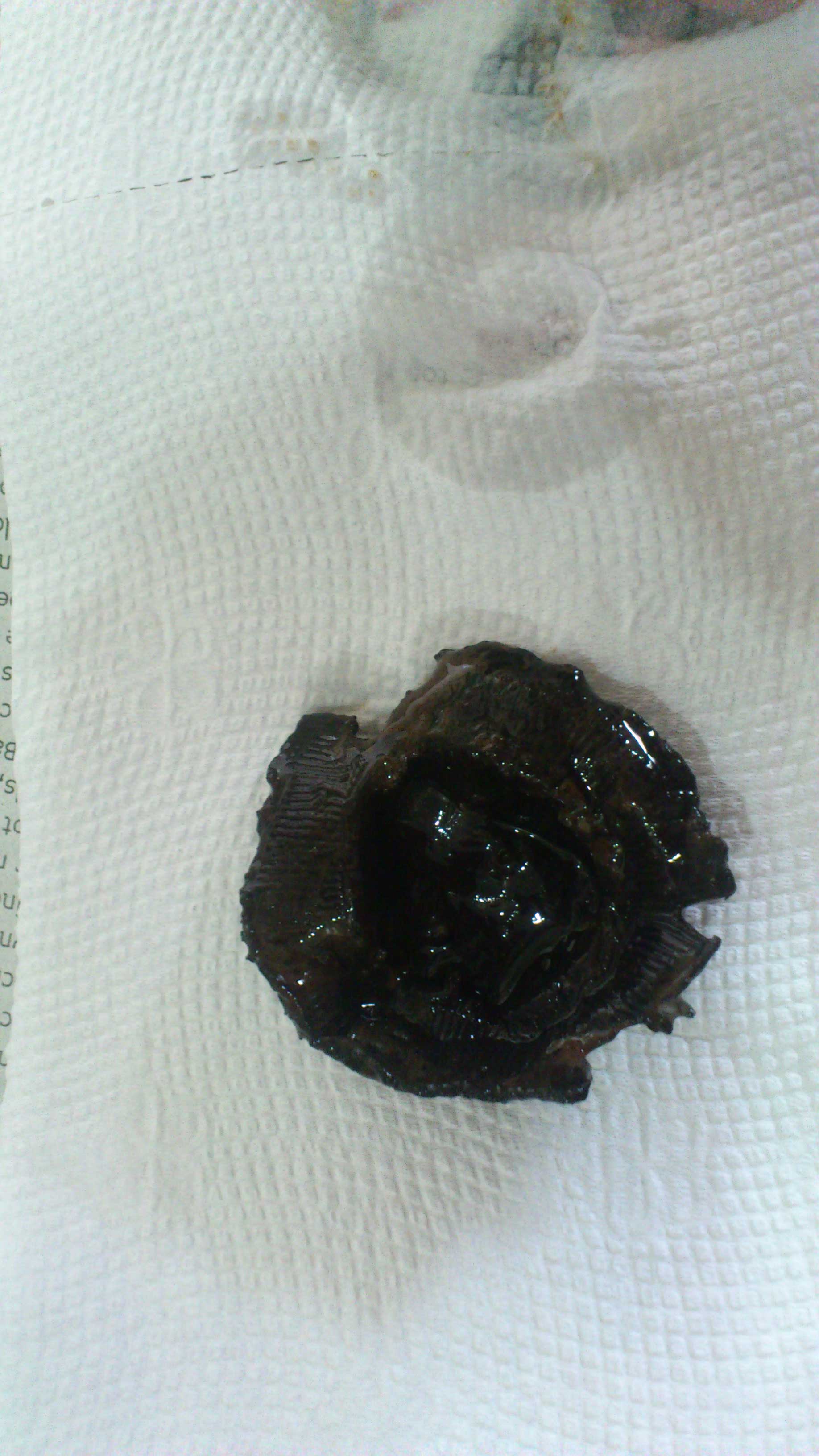
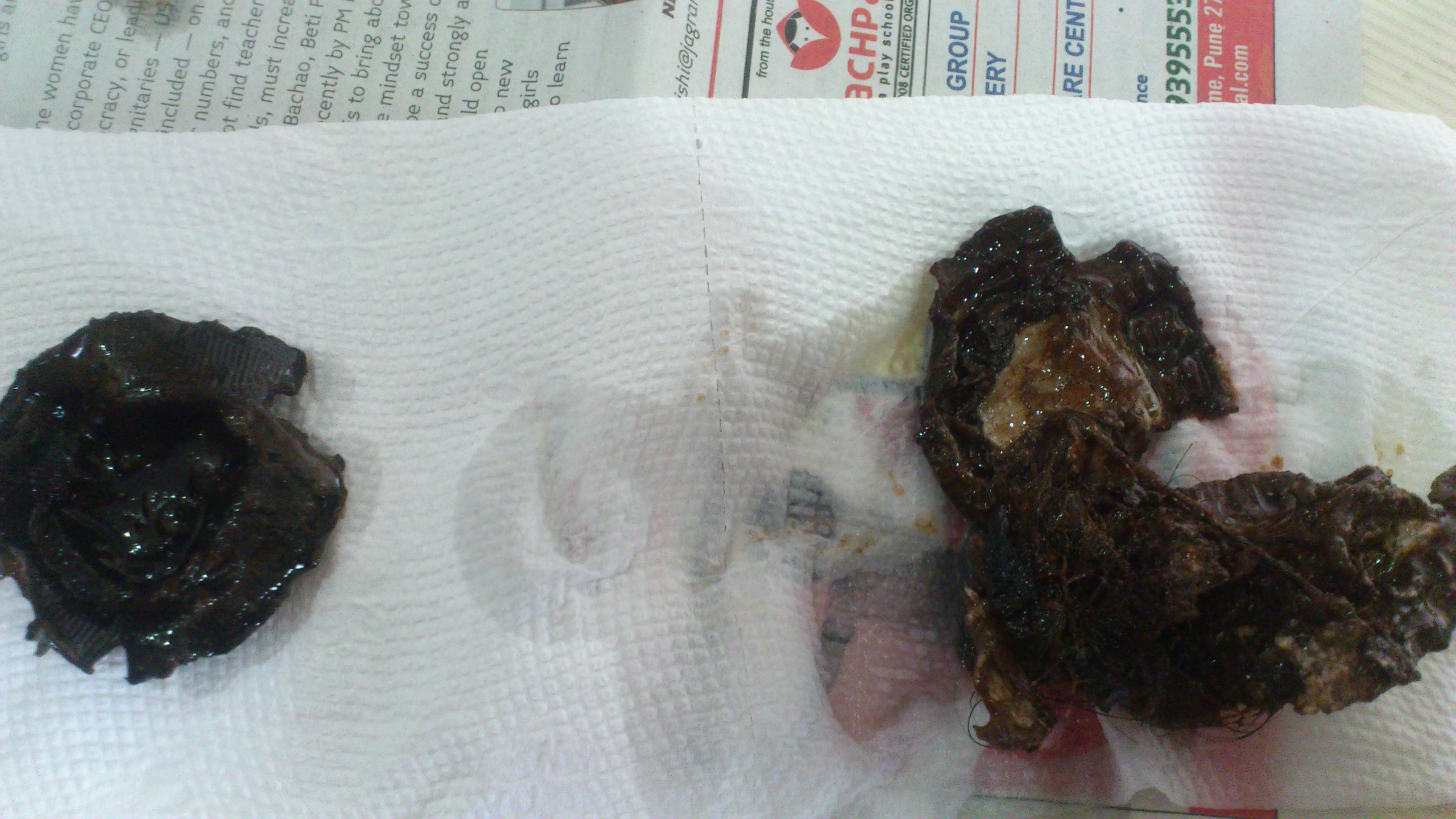
Stapler pins in abdomen engulfed by dog